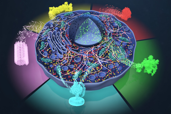
Researchers working to understand and develop better therapies for diseases like HIV, cancer, and Parkinson’s have more power than ever before to “see” proteins and their interactions inside cells. Cryogenic electron microscopes visualize frozen proteins in their near-native state, capturing ultra-detailed images. Artificial intelligence-powered analysis then detects patterns and produces accurate 3D models.
But the process of turning raw data into usable protein structures requires stitching together multiple software tools and can take months.
Now, Duke researchers have developed a user-friendly, web-based software tool that cuts that time down to mere days. Called nextPYP, the platform automates many of the most time-consuming steps, using advanced machine learning to streamline data analysis, improve storage efficiency, and accelerate processing.

In a study published this month in Nature Protocols, Alberto Bartesaghi, PhD, associate professor of computer science and biochemistry, and colleagues demonstrate how nextPYP can be used to determine the structure of key proteins, including HIV-1 Gag and human ribosomes, directly within cells.
Bartesaghi said the tool could be a game-changer. “By enabling more efficient and precise mapping of these challenging targets within their native environments, nextPYP could significantly accelerate drug discovery and deepen our understanding of a wide range of diseases.”
“Before nextPYP, there was no unified, accessible, end-to-end solution that combined advanced AI-driven data processing with high-resolution structural refinement,” he said.
Future research from this team will focus on using the tool to study a broader range of proteins, including membrane proteins, which play a critical role in many cellular processes and are estimated to be the targets of nearly 70 percent of approved therapies.
Funding: The National Institutes of Health and the Chan-Zuckerberg Initiative.