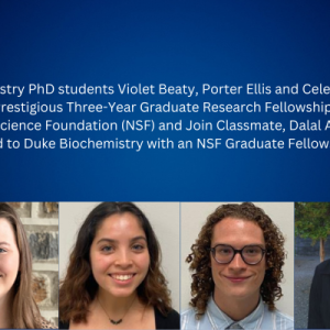
Three PhD Students Awarded Three-Year Graduate Research Fellowships from the NSF
Biochemistry PhD students Violet Beaty, Porter Ellis and Celeste Marin have been awarded prestigious three-year Graduate Research Fellowships from the National Science Found
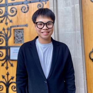
Bach Nguyen Awarded Graduate/Professional Academic Exemplar of the Year from DISC
With a population of nearly 3,000 on campus, international students are an integral part of Duke’s community.

Congratulations to Skyler Cochrane, who has been invited for an oral presentation at the upcoming ASM meeting in Atlanta!
Congratulations to Skyler Cochrane, who has been invited for an oral presentation at the upcoming ASM meeting in Atlanta!

Emily Cannistraci Awarded the Jo Rae Wright Fellowship
The Jo Rae Wright Fellowship for Outstanding Women in Science was created in loving memory of Jo Rae Wright, former dean of the Duke University Graduate Sc
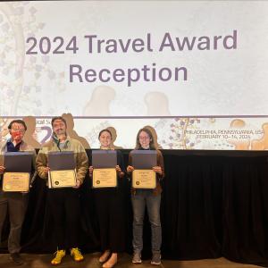
Four Biochemistry Students Receive Travel Awards to Attend this year's Annual Biophysical Society Meeting
Four Duke Biochemistry graduate students, Aaron May, Gus Lowry, Emily Cannistraci and Ruth Parsons, have been awarded travel awards to attend this year’s Annual Biophysical Societ

Duc Huynh Receives 2024 SoM Dean's Award for Research Excellence (DARE)
Congratulations to Duc Huynh, who has received the 2024 SoM Dean's Award for Research Excellence (DARE)!

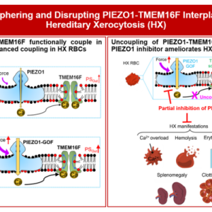
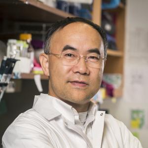
Dr. Pengfei Liang Awarded 2-Year Postdoctoral Fellowship from the American Society of Hematology
Congratulations to Dr. Pengfei Liang from the Yang lab, who won the prestigious 2-year postdoctoral fellowship from the American Society of Hematology.